August 13, 2024
Purpose of the Study
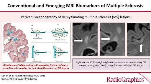
The purpose of this study was to determine how often new lesions appear in the cervical spinal cord without any accompanying new lesions in the brain on MRI in patients with multiple sclerosis (MS). The study focused on this specific spinal cord segment due to its frequent involvement in MS and its common inclusion in routine monitoring alongside brain imaging. Additionally, the study aimed to identify clinical and MRI factors linked to the development of isolated cervical spinal cord lesions and to assess whether these lesions correlate with future relapses or disability progression in MS patients.
Who Performed the Study
The study was a collaborative effort conducted by researchers from Toronto Radiology at St. Michael’s Hospital, the Department of Medical Imaging at the University of Toronto, and the Barlo MS Centre at St. Michael’s Hospital, Unity Health Toronto. The lead researchers were Dr. Aditya Bharatha, a staff radiologist, and Dr. Jiwon Oh, a staff MS neurologist. Key collaborators included Drs. Timothy Reynold Lim and Sunitha Kumaran, neuroradiology fellows; Drs. Suradech Suthiphosuwan and Amy Lin, staff radiologists; and Dr. Adrian Espiritu and Ashley Jones from the Barlo MS Centre, St. Michael’s Hospital, Unity Health Toronto, and Division of Neurology, Department of Medicine, University of Toronto.
How the Study Was Done
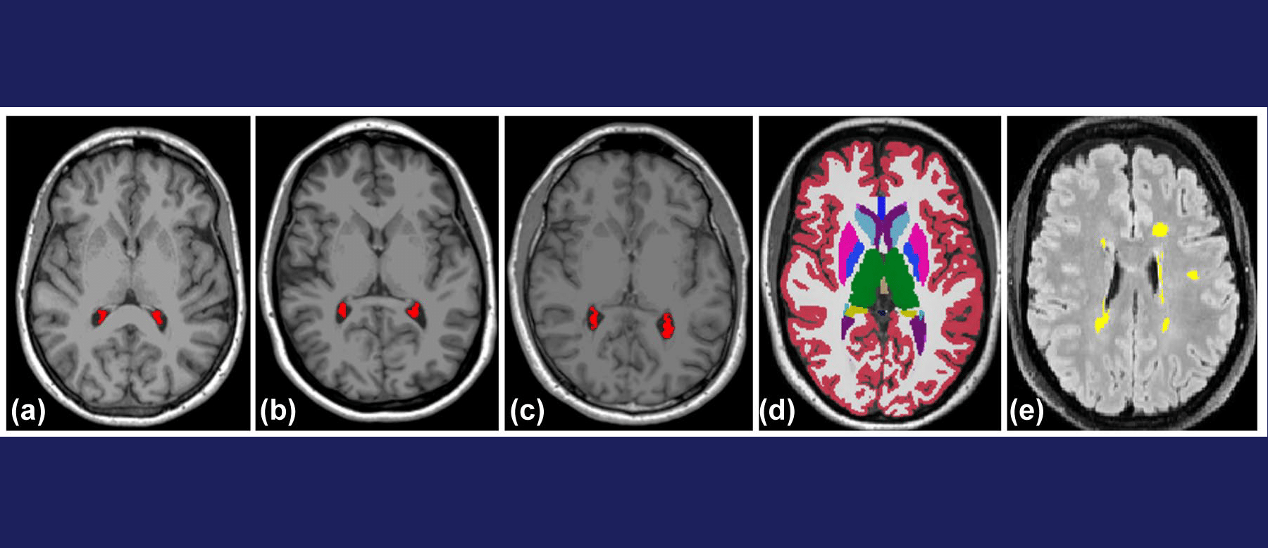
This retrospective study analyzed 1,576 MS patients who underwent follow-up 3 Tesla MRI of the brain and cervical spinal cord over 9 months. The MRI scans were reviewed for the presence of new brain lesions and cervical spinal cord lesions. Clinical records were also reviewed to document any relapses between sequential MRI scans and to track clinical relapses or disability progression following the follow-up MRI.
Key Findings
The study found that less than 2% of clinically stable MS patients developed new lesions isolated to the cervical spinal cord while having no symptoms over a median follow-up period of 13-14 months. The occurrence of these lesions was associated with the presence of three or more new brain lesions. Importantly, these spinal cord lesions, when considered independently of new brain lesions, did not increase the risk of future relapses or disability progression over a subsequent 26-month period. These findings suggest that routine cervical spine MRI may not be necessary for monitoring clinically stable MS patients if they are already undergoing advanced brain imaging with state-of-the-art 3 Tesla MRI and 3-dimensional volume subtraction techniques.
Summary of the Full Citation
For more in-depth information, refer to the full citation into the necessity of routine spinal cord MRI for MS monitoring.